new: mike tutors and offers personalized advice for the usmle (click here for more info)
Showing posts with label Microbiology. Show all posts

Sunday, May 6, 2007

The Tuberculous Granuloma is High Yield for the USMLE

Ahh the Granuloma - a great example of topic that overlaps multiple subjects and any topic that brushes borders with the likes of pathology, immunology and microbiology make for great questions. Here are 5 steps to the granuloma in perfect 3rd grader doodle form:


Step 1:
Inhale the red, ACID FAST rod


M. tuberculosis is only acid fast why?

B/c of the MYCOLIC ACID in the cell wall resists decolorization with acid-alcohol and so it remains red which is the color of the initial stain, carbol fuchsin.

What else stains acid fast? Nocardia which is “partially acid fast”


Step 2: Phagocytosis by alveolar macrophages

Circulating monocytes roll on the vascular endothelium (d/t selectins) and adhere to it (d/t ICAMs) and then transmigrate into the affected area where they are called tissue macrophages.

Macrophages are the main players in the formation of a granuloma

Step 3: Antigen Presentation


Step 4: T Cell Activation


CD4 T Cells are involved

The TH1 subtype

No CD4 T Cells? No Granuloma. AIDS patients who loose their CD4 t-cells well before loosing their CD8 T Cells will make very weak if any granulomas.


Step 5: Macrophage Activation


Did I mention what the main player of a granuloma is? Oh yea, its the macrophage.

The Caseous Granuloma


Note that the caseous necrosis is cottage cheese like and is due to the destruction of the M. TB organism's cell wall's mycolic acids (mycolic acids are lipids).

Thursday, May 3, 2007

Viruses are High Yield for the USMLE Step 1

I suggest when trying to memorize viral structures make a priority list. Common viruses and ones that also overlap with pathology are the ones to focus on here since they can be asked about in many different ways.

Knowing DNA vs RNA is the most important distinguishing feature. Furthermore, f you know if its DS or SS you'll probably get the points.

Enveloped vs Naked: If you can memorize enveloped vs. naked for each virus then god bless, but I like this:
Envelopes are made of a lipid bilayer which are prone to destruction by the environment whereas naked viruses are more resistant to environmental stresses.

A 46 year old female who has genital warts (d/t HPV) takes a shower in her home during which time the virus sheds on the floor. Later her child presents with a wart on his toe.
This all happened since this virus had its tough nucleocapsid for protection and not a wimpy envelope. Take home message is if the virus is capable of fomite transmission it is probably naked.

Play odds, especially for rare viruses with less of a chance of being asked. I walked into the exam knowing that if I saw an RNA virus that looked deadly or pretty rare I would guess that it is SS (-) linear w/envelope.

Here is my list of High Yield Viruses and why I think they have a great chance of being asked
  • Herpesviruses - DS linear DNA
    • this family includes so many viruses that overlap with pathology I list it first since I think this is most important.
  • All the viruses that cause hepatits - see First Aid for them since these are important!
    • Note that Hep B carries the reverse transcriptase enzyme with it just like the retroviruses do.
  • HIV - SS (+) linear RNA
    • If you don't get at least 1 question on AIDs I will give you my blog. Seeing if you know virus structure is a possible secondary question.
  • Papovavirus - DS (-) linear
    • besides HPV causing cervical cancer don't forget about molluscum contagiosum which causes the umbilicated wart (center of wart is depressed like an "innie" belly button).
  • Paramyxoviruses - SS (-) linear RNA with helical capsid symmetry
    • this family is responsible for causing croup (parainflunza virus), bronchiolitis (d/t RSV), measles and mumps.
  • Parvovirus - SS linear DNA
    • It, along with Hepadnavirus, is exception to the rule that all DNA viruses are linear.
    • It overlaps with pathology since it causes aplastic anemia in patients with sickle cell disease
      • Pt might give you a history of having recent contact with a kid who had a slapped cheek appearance.
      • Blood labs will give you a low RBC count with low reticulocytes (less than or equal to 3% of hematocrit - I wouldn't freak out about not knowing how to correct a reticulocyte count).
  • Influenza virus (an orthomyxovirus) - SS (-) linear segmented RNA
    • the NBME and practice questions I've seen love the fact that this genome is segmented since:
      • Genetic shift - if our influenza virus recombines with a pig's or chickens genome we get pandemics - we're all screwed (small minor mutations lead to drifts causing an epidemic in a much smaller area w/ much less people). involved)
      • If you digest the genome and view it with northern blotting you will see each of these (7 or 8) segments as separate bands.

Wednesday, April 18, 2007

Burkitt's Lymphoma is High Yield for the USMLE Step 1

Burkitt Lymphoma
a form of non-Hodgkin's lymphoma

Associated with Epstein Barr Virus and is commonly located in the jaw of Africans (the classic patient)

Classic Translocation = t(8;14) which moves the c-myc gene on chromosome 8 right next to the Immunoglobulin (Ig) Heavy Chain.

Here is a joke for you. I hope you find it hilarious. Okay, a giggle will do. Tell it to someone else, embrace the awkwardness that ensues and that should tattoo it to your brain for a while.

How come Mick ate one for?

-----Here is what the joke really means----
come Mick = c-myc
ate = 8 - since c-myc is on chromosome 8
one = 1
for = 4

To get heavy!
heavy = Ig Heavy chain which is on chromosome 14


And here's a little doodle to drive it on home:


That big guy there is Mick (myc) who has just ate (8) one (1). For (4) what? To get heavy.
c-myc is on chromosome 8
Ig Heavy chain is on chromosome 14

Believe me, this joke is much funnier if you're telling it to yourself when you are looking at a question on it.

Here is what some practice questions I've seen ask and is mentioned in various review therefore I made sure I knew this for my exam although I had to look it up again for this post.

The function of c-myc: c-myc is a proto-oncogene which codes a protein that regulates nuclear transcription. When it is moved next to the gene for the Ig heavy chain it becomes constantly active since the body is constantly making Ig heavy chains. At this point it is now called an oncogene. c-myc now causes the B-cells to constantly reproduce themselves over and over again. This results in a high grade lymphoma

This is in contrast to B-cell follicular lymphomas in which BCL-2 (a gene which promotes apoptosis). When BCL-2, a tumor suppressor gene, is translocated it becomes functionally inactive and so B-cells can't undergo apoptosis the same anymore. This is an example of low grade tumor.

So I think this is how you should think about it: Burkitt's lymphoma is when B-cells actively divide whereas in B-cell follicular lymphomas, B-cells don't die.

Also, here's the classic"Starry Sky Appearance"
(source: pathguy.com)

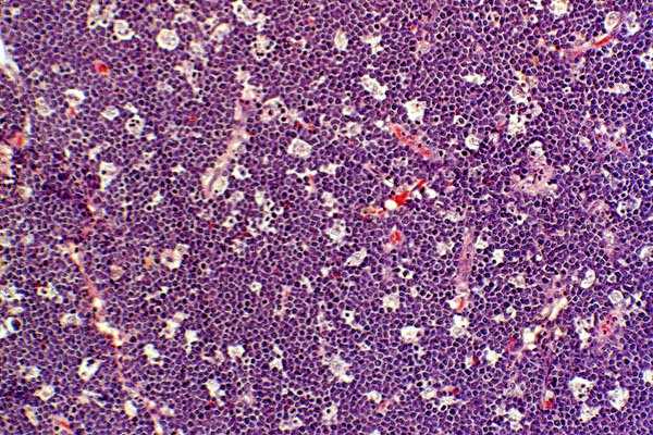
I just want to point out here what that refers to. The dark purple areas are really just a bunch of neoplastic B-cells that are dividing much more rapidly now that c-myc is always active. ("Dark is Dividing") The stars are the lighter areas which are much fewer in number - these are the macrophages. I'm not sure what they are doing there, but I do know that they are NOT the problem. Perhaps they are trying to help out in getting rid of the tumor.

That's what I think is really important for this tumor that seems to be a pretty hot topic for the exam.

Thursday, March 29, 2007

Tabes Dorsalis


  • This is a manifestation of tertiary (late) syphilis called neurosyphilis
    • caused by infection with treponema pallidum, a spirochete
    • tertiary syphilis also turns the vasa vasora (blood vessels that supply other blood vessels, eg aortic root) into a bunch of granulomas →:
      • Aortic regurgitation – know how to recognize!
        • Diastolic decrescendo murmur heard best along the left sternal border
      • Aneurysm of the ascending aorta
  • Note that tabes dorsalis only affects the dorsal horns.
  • This patient had a positive (+ve) Rhomberg's sign
    • Rhobmerg's sign
      • Patient's balance is fine with his eyes open
        • Visual input supplies spatial awareness/proprioception
      • Upon closing his eyes the pt sways
        • loss of proprioception from dorsal column

(If patient is swaying with eyes open and closed, this is likely a lesion in the cerebellum or vestibular system (eg. CN VIII) and would NOT qualify as a +ve Rhomberg's sign)

  • Can you guess the level from which this spinal cord section was taken
A) C1
B) C4
C) T4
D) L4
E) S1

Answer posted on 4/2/07

Note the lateral horns (sometimes called intermediolateral cell column):
(I have circled the right one and have drawn an arrow to the left one)
This area of white matter contains preganglionic sympathetics which arise from the hypothalamus and will synapse onto a postganglionic sympathetic fiber in the paravertebral (aka chain) ganglion. Question for you... would the neurotransmitter used here be
a) norepinephrine (Norepi)
b) epinephrine (Epi)
c) acetylcholine (ACh)
d) glycine (Gly)
e) glutamate (Glu)
f) seratonin (5-HT)

Answer is c) Ach
Remember, all preganglionic autonomic fibers use ACh
Epinephrine is released from the adrenal medulla when preganglionic sympathetic fibers release ACh onto it.

Now, back to the level of the spinal cord:
Since we are talking about sympathetic (aka thoraco-lumbar) outflow, you should know right away that this slide must therefore have been sliced from somewhere between T1-L2.
Knowing this would have gotten you the right answer to my question.

If the question writer is out for your blood he may ask you to distinguish between 2 choices within the range of the lateral horn. Notice the shape of the above section. Now compare it to an upper lumbar section. See the difference? The lumbar section has only one dorsal column and it is just a lot more round. That "just a lot more round" part (I bet) is enough to differentiate for purposes of the exam. But feel free to knock yourself out, I know I did.


Epstein Barr Virus

  • Attaches and activates B-lymphocytes CD21 (CR2) receptor
    • These activated B cells promotes T cell proliferation
    • The effected B-lymphocytes are NOT what are detected by the monospot test.
  • Atypical lymphocytes (“Downy Cells”)
    • Detected by the monospot test
      • AKA (heterophile antibody test)
    • Are CD8+ T lymphocytes
  • Associated with:
    • Heterophile positive infectious mononucleolus
    • Burkitt's Lymphoma
    • Nasopharyngeal Carcinoma
  • Structure
    • a "HAPPy" double-stranded linear DNA virus (see First Aid virus section)